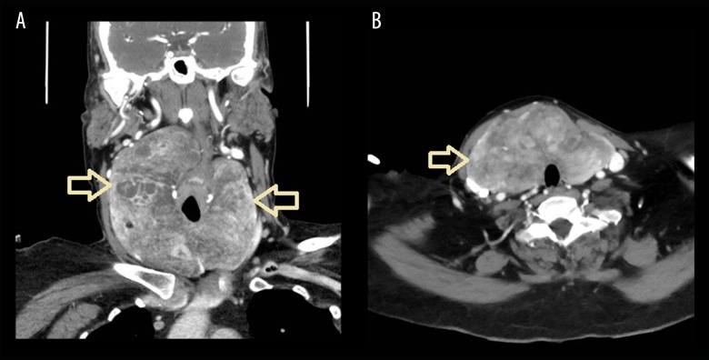
Figure 1.
(A) A coronal CT with contrast showing heterogeneous enhanced thyroid with cystic changes and a heterogeneous enhanced thyroid mass lesion. The thyroid gland extends up to the pharynx and down to the sternum (arrows). (B) An axial CT with contrast showing heterogeneous thyroid with a heterogeneous enhanced mass lesion. The thyroid extends up and posteriorly reaching the right pyriform fossa, exerting compression on the oropharynx and extending down the retrosternal (arrow). The tracheal air column in the neck is patent and displaced to the left side.

