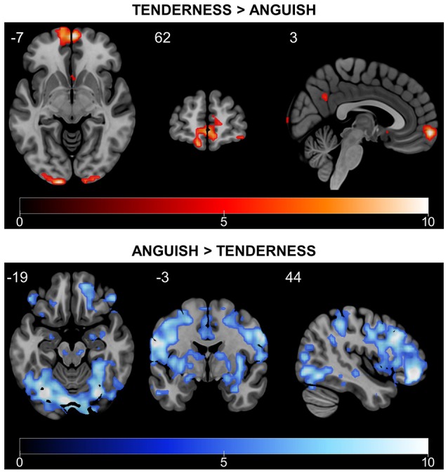
Figure 6.

Differential rtfMRI-NFB-related brain responses for tenderness and anguish conditions. Tenderness vs. anguish rtfMRI-NFB recruited the septo-hypothalamic area, the frontal pole and the precuneus. Anguish vs. tenderness rtfMRI-NFB recruited a more widespread network including the superior/middle frontal cortex, frontal pole, parietal cortical regions, temporal regions (middle and inferior) and other regions (lateral occipital, central operculum, cerebellum). Results were estimated across all participants (N = 16, two scans per subject) via fixed-effect analysis and whole-brain FWE correction with p < 0.05, T > 4.716).
