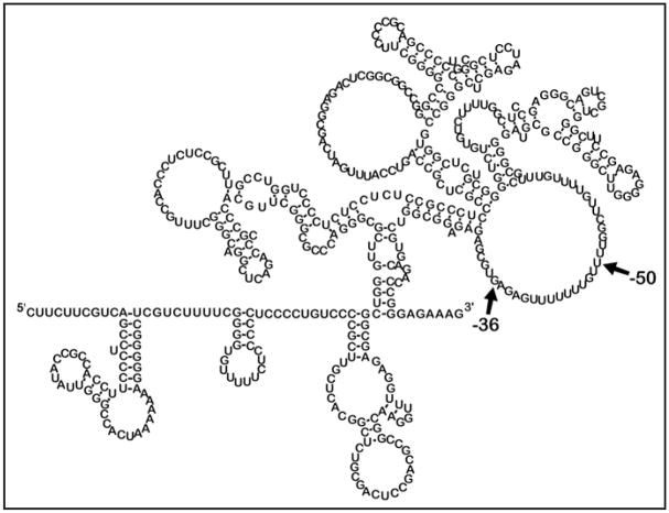
Figure 4.
MFOLD secondary structure prediction for the 472 nucleotide p27Kip1 5′-UTR. Banding patterns seen after cleavage/RT (as in Fig. 3) were used to identify areas of single- and doublestrandedness of the p27Kip1 472 nucleotide 5′-UTR. Based on these input parameters which forced or prohibited pairing, the MFOLD application predicted this complex secondary structure. The 5′ and 3′ ends of the 5′-UTR are indicated. The region between -36 and -50 indicate the proposed ribosome entry window for IRES-mediated initiation of translation (see Fig. 5).

