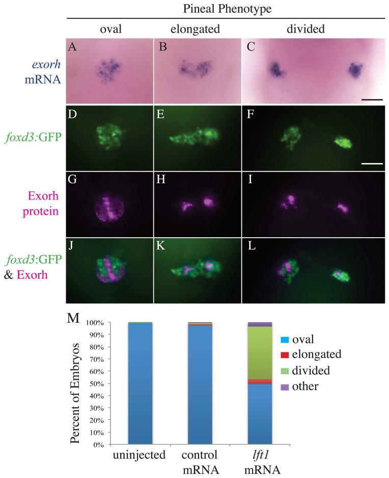
Figure 2. Projection neuron and photoreceptor differentiation occurs in the pineal glands of Lft1 overexpressing embryos with open neural tubes.
(A – C) The photoreceptor specific gene exorh is expressed in embryos with all three pineal phenotypes (n = 21 oval, n = 2 elongated, n = 6 divided). (D – L) Embryos were analyzed for both foxd3:GFP transgene expression and Exorh protein expression, and a representative embryo with an oval pineal (D, G, J), with an elongated pineal (E, H, K), and a divided pineal (F, I, L) are shown. (D–F) are images of the foxd3:GFP expression, (G–I) are images of the Exorh immunostaining, and (J–L) are overlays of the two expression patterns. Note that in (J–L), foxd3:GFP and Exorh are expressed in distinct regions of the pineal, suggesting the foxd3:GFP transgene is primarily found in pineal projection neurons at this stage of development (n = 36 oval, n = 4 elongated, n = 11 divided). (M) Percentage of lft1 mRNA injected embryos expressing the foxd3:GFP transgene in an oval pattern (closed anterior neural tube), or elongated or divided pattern (open neural tube). n > 120 embryos. All embryos were at 48 hpf. All images are dorsal views, anterior to the top. Scale bar = 25 μm.

