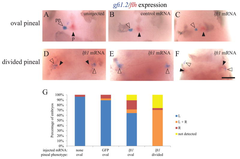
Figure 6. Both the left and right sides of the brain form a parapineal when the epithalamus is divided.
foxd3:GFP homozygous embryos were injected with GFP mRNA or lft1 mRNA at the one to two cell stage. Injected embryos and their uninjected siblings were sorted by the pineal phenotype using fluorescent microscopy, fixed at 3 dpf, and then assayed for gfi1.2 in the parapineal (pp, closed arrowheads) and flh in the pineal (p, open arrowheads). (A–C) In uninjected embryos, embryos injected with control GFP mRNA, and in lft1 mRNA injected embryos with oval shaped pineals, the parapineal organ is located adjacent to left side of the pineal. (D–F) In the lft1 injected embryos with divided pineal organs (not shown, as pineal is out of the focal plane of the image), there is a region of gfi1.2 expression on both the left and right sides of the brain, suggesting two parapineal organs have formed. (G) Graph of data represented in A–F. The experiment was repeated four times and representative images are shown. n ≥ 26 for each pineal phenotype. Dorsal views, anterior to the top. Scale bar = 50 μm.

