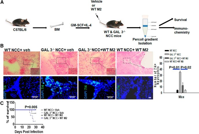
Figure 7.
Galectin-3-expressing M2 macrophages exhibit protective role during NCC infection. A, Schematic representation of the adoptive transfer experiment as described in Materials and Methods. Purified bone marrow macrophages from WT mice were differentiated to M2 phenotype (WT M2) by exposure to IL-4 in vitro. WT (WT NCC) or Galectin-3−/− (GAL3−/NCC) mice, undergoing NCC due to intracranial infection with M. corti, received WT M2 (WT NCC + WT M2; GAL3−/−NCC + WT M2) or vehicle (WT NCC + Veh; GAL3−/NCC + Veh) at 1 and 2 weeks after infection Disease severity among these four groups was compared at 3 weeks of NCC. B, Top, Representative images of H&E-stained brain cryosections. Bottom, IF microscopy images of brain cryosections to compare accumulation of 7/4+ (green) neutrophils. Nuclei (blue) were stained with DAPI. Bar graph represents 7/4+ neutrophils manually counted from images shown at bottom from 3 independent experiments with 3 mice per experiment. p values (t test). C, The survival of WT NCC and Galectin-3−/− NCC mice adoptively transferred with WT-M2 macrophages or vehicle was monitored for 3 weeks. Statistical significance was determined by Student's t test. p values (t test and Log-rank [Mantel–Cox] Test). Migration of CFSE-labeled WT-M2 cells (F4/80+CFSE+) into the CNS of the recipient WT and Galectin-3−/− NCC mice at 24 h postadoptive intravenous transfer is presented in Figure 7-1.

