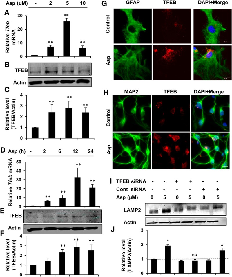
Figure 2.
Aspirin stimulates lysosomal biogenesis via upregulating TFEB. A–C, Mouse primary astrocytes were treated under serum-free conditions with different doses of aspirin, followed by monitoring of Tfeb mRNA expression at 8 h (A) and protein levels of TFEB at 24 h (B) of treatment and subsequent densitometric analysis of TFEB protein expression (C). Data are shown as fold change (mean ± SD) with respect to untreated control. D–F, Time point analysis for TFEB expression with 5 μm aspirin treatment by monitoring mRNA levels by quantitative RT-PCR (D), protein expression by immunoblot (E), and densitometry of TFEB protein levels (F). Data are shown as fold change (mean ± SD) with respect to untreated control. G, Mouse primary astrocytes treated with 5 μm aspirin for 24 h, followed by double labeling with TFEB and GFAP. Scale bar, 20 μm. H, Primary cortical neurons were treated under the same conditions and stained for TFEB and neuronal marker MAP2. Scale bar, 10 μm. I, J, Primary astrocytes were transfected with Tfeb siRNA or scrambled siRNA for 48 h and subsequently treated with 5 μm aspirin for 24 h, followed by Western blot (I) and densitometric (J) analysis for LAMP2 protein levels. All data are shown as mean ± SD. Statistical analysis was conducted using Student's t test; ns, nonsignificant; *p < 0.05, **p < 0.001.

