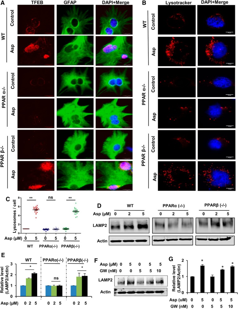
Figure 4.
Induction of aspirin-mediated lysosomal biogenesis in WT and PPARβ−/− astrocytes, but not in PPARα−/− astrocytes. A–E, Primary astrocytes from WT, Ppara-null, and Pparb-null mice were treated with 5 μm aspirin for 24 h, followed by immunofluorescence staining for TFEB and GFAP (A; scale bar, 20 μm). Nuclei were stained using DAPI. B, Live cell staining using LysoTracker Red. Scale bar, 10 μm. C, Quantification of the lysosome number per cell using ImageJ. Data are shown as mean ± SEM of fold change relative to the untreated WT control. Twenty images per condition from three independent experiments were quantified. Immunoblot analysis for LAMP2 (D) and densitometric analysis of LAMP2 protein levels (E). Data are shown as mean ± SD for fold change with respect to untreated control in each group. F, G, WT astrocytes were pretreated with 5 and 10 nm PPARγ inhibitor GW9662 for 1 h, followed by 24 h treatment with 5 μm aspirin and analyzed for LAMP2 protein levels by Western blot (F) and densitometric analysis (G). Data are shown as mean ± SD for fold change normalized to the untreated control. All statistical analyses were done using Student's t test; *p < 0.05, **p < 0.001.

