Figure 5.
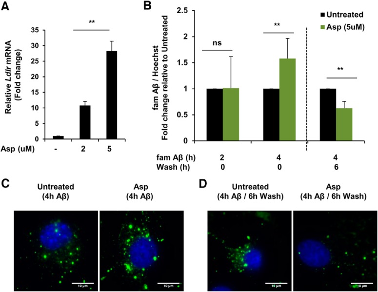
Aspirin enhances uptake and degradation of Aβ42 in mouse primary astrocytes. A, Primary astrocytes were treated with 5 μm aspirin for 4 h, followed by monitoring the mRNA expression of Ldlr by quantitative RT-PCR. Data are shown as fold change (mean ± SD) with respect to untreated controls. B, Treatment for 24 h followed by FAM-Aβ(1–42) uptake and degradation assay. Cells were incubated in medium containing 500 nm FAM-Aβ for 2 h, 4 h for monitoring the uptake. For degradation, the cells were grown in Aβ-free regular medium for an additional 6 h. Data are shown as fold change (mean ± SD) with respect to untreated control. C, Treatment for 24 h followed by 4 h incubation in medium containing HiLyte Fluor 647-Aβ for monitoring Aβ uptake. D, For degradation, the cells were washed in medium for an additional 6 h. DAPI was used to stain the nuclei. All statistical analyses were performed using Student's t test; **p < 0.001.

