Figure 7.
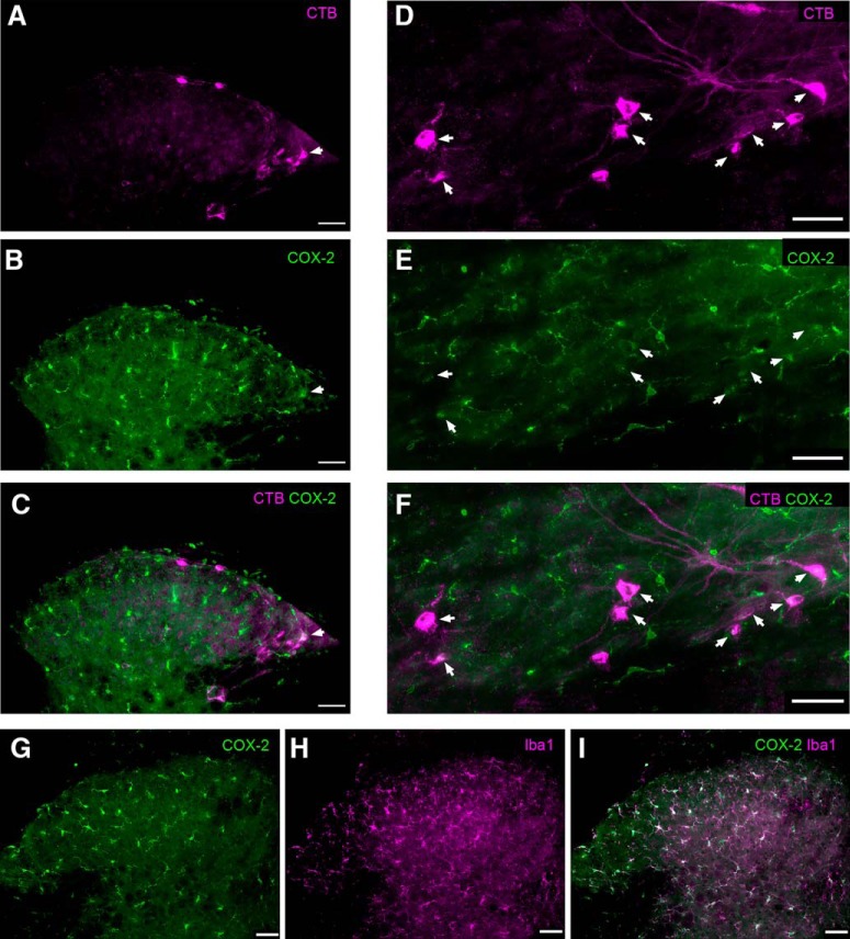
Constitutive expression of Cox-2 in the adult mouse SDH. A, Transverse spinal cord section showing the ascending projection neurons that were identified via retrograde transport of CTB previously injected into the parabrachial nucleus. B, Same section as in A, illustrating immunoreactivity (IR) for Cox-2. Arrowhead indicates a CTB-labeled projection neuron that exhibits Cox-2 IR. C, Merged image (from A and B) demonstrating the relative distribution of CTB (magenta) and Cox-2 IR (green) in the SDH. The colocalization of CTB and Cox-2 (white) suggests Cox-2 expression within a subset of spinal projection neurons (arrowhead). D–F, Horizontal sections of the SDH illustrating additional examples of projection neurons back-labeled with CTB (D) that also possess Cox-2 IR (E), as evidenced by the colocalization seen in F (arrowheads). G–I, Nonetheless, the majority of Cox-2 IR in the SDH (green) was colocalized with the microglial marker Iba1 (magenta). Scale bars, 50 μm.

