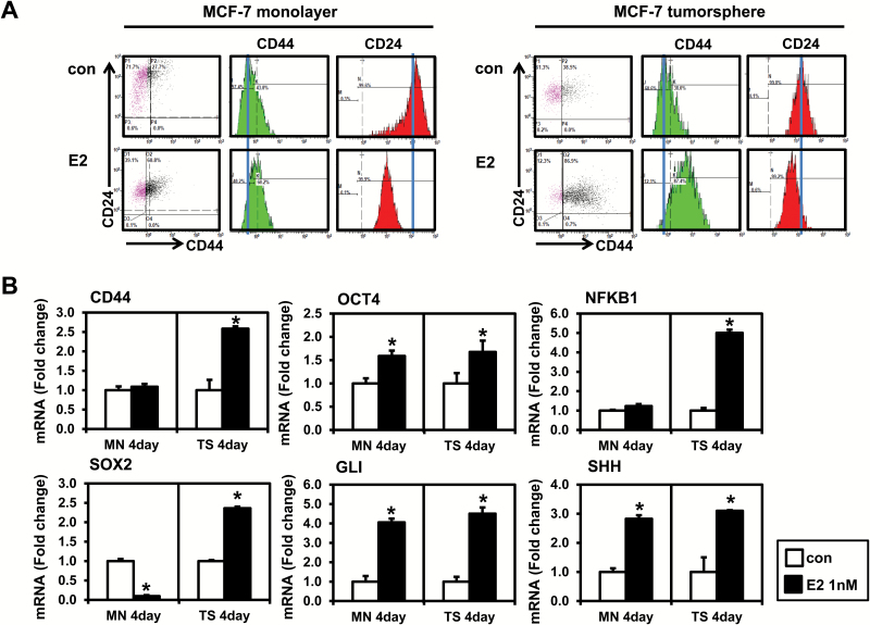
Figure 1.
Effects of estrogen (E2) on the CD44/CD24 expression and the level of stem cell markers in MCF-7 monolayer cells and tumorspheres. (A) MCF-7 monolayer cells were plated at a density of 8 × 10⁴ cells/ml in six-well plates and treated with the vehicle control (con) or E2 (1 nM) for 4 days. MCF-7 tumorspheres were formed by plating 10000 cells/ml in ultra-low attachment six-well plates and treated with E2 (1 nM) for 4 days. Cells were stained with combinations of antibodies against CD24 and CD44, and then, flow cytometry was performed. Experiments were performed in triplicate, and representative histograms from flow cytometry are shown. Histograms show the mean fluorescence intensity of CD44 (PE-Green) and CD24 (FITC-Red). (B) qPCR analysis was performed on MCF-7 monolayer cells and tumorspheres treated with E2 and analyzed for markers of cancer stem cells. The data are represented as mean ± standard deviation (SD). Here, n = 3 represents independent experiments, *significantly different from the respective control (P < 0.05). Cycle numbers for genes related to CD44, OCT4, NFKB1, SOX2, GLI and SHH for control group in MCF-7 monolayer cells were 25, 25, 24, 31, 34 and 28 and for control group in MCF-7 tumorspheres cells were 25, 28, 26, 30, 35 and 30, respectively.

