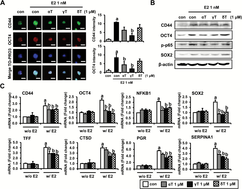
Figure 4.
Effects of tocopherols on expression of E2-target genes and stem cell markers in E2-treated MCF-7 tumorspheres. (A) Immunofluorescence analysis of was performed on MCF-7 tumorspheres collected from 4 days of treatment with control, E2 (1 nM) or tocopherols (1 μM). MCF-7 tumorspheres were fixed using 4% paraformaldehyde and stained with antibodies against CD44 (green) and OCT4 (red). Nuclei were stained with TO-PRO3 (blue). Representative pictures are shown, scale bars: 200 μm. The quantification of CD44 and OCT4 level by ImageJ program from three independent experiments was shown. a,bSignificantly different from the control and E2 control, respectively (P < 0.05). (B) Western blot analysis was performed on MCF-7 tumorspheres collected from 4 days of treatment with control, E2 (1 nM) or tocopherols (1 μM) and analyzed for markers associated with stem cell signaling. β-Actin was used as a loading control. (C) qPCR analysis was performed on tumorspheres collected from 4 days of treatment with control, E2 (1 nM) or tocopherols (1 μM) and analyzed for markers associated with stem cell-related genes, including CD44, OCT4, NFKB1 and SOX-2, and E2-related genes, including TFF, CTSD, PGR and SERPINA1. The data are represented as mean ± SD. Here, n = 3 represents independent experiments, a,bsignificantly different from the control and E2 control, respectively (P < 0.05). Cycle numbers for genes related to CD44, OCT4, NFKB1, SOX-2, TFF, CTSD, PGR and SERPINA1 for control group were 25, 27, 26, 30, 18, 20, 30 and 32, respectively.

