
Fig. 2.
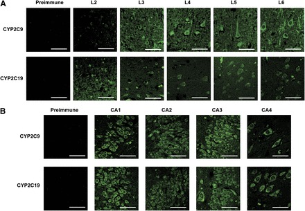
(A and B) Immunohistochemical detection of CYP2C9 and CYP2C19 expression in the human frontal cortex (A) and hippocampus (B). Negative controls incubated with preimmune sera for CYP2C9 and CYP2C19 were performed with layers 5 and 6 (A) and CA4 (B), respectively, and were representative of other layers. (A) CYP2C9 and CYP2C19 proteins were detected in layer 2 through layer 6 of the frontal cortex, predominantly in the soma of neuronal cells. Expression of CYP2C9 was also observed in neuronal axons and dendrites, predominantly in layers 5 and 6. (B) CYP2C9 and CYP2C19 were both located throughout the hippocampus; however, expression appeared to be highest in the CA1 region in both cases. In addition, CYP2C9 was also highly expressed in the CA3 region. Bar, 50 μm.