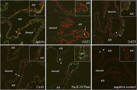
Fig. 5.

Immunolocalization of Na,K-ATPase, Cx43, MRP4, OAT1, and OAT3 in paraffin-embedded human eye. Na,K-ATPase was used as a marker of basolateral membranes of pigmented and nonpigmented cells. Cx43 was used as a marker for the apical membrane of both cell types. Proteins of interest are in green and nuclei are stained red with propidium iodide. The arrowheads point to pigmented cells whereas the arrows point to nonpigmented cells. The insets are magnifications of the areas near the arrow and arrowhead. Limited immunoreactivity was detected when the primary antibody was omitted (negative control). AH, aqueous humor.
