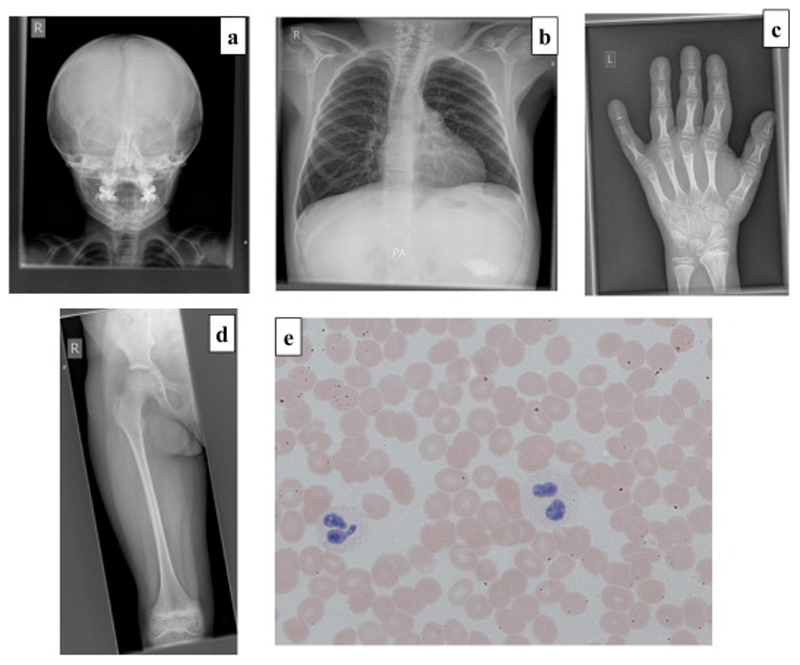
Figure 2.
a-d: Radiographs demonstrating slender ribs, tubular long bones with thin cortices and osteopenia consistent with a diagnosis of OI. 2a: AP skull radiograph (aged 6 years) There are multiple Wormian bones; the clavicles are slender and the erupted teeth are relatively dense. The anterior fontanelle remains open.
2b-d: Selected images from a full dysplasia skeletal survey (aged 9 years 8 months)
2b: Left hand. There is significant periarticular osteopenia (see Fig 2c). The metacarpals (and less marked) the proximal phalanges are overmodelled and there is an ivory epiphysis of the terminal phalanx of the fifth finger. The terminal tufts are prominent.
2c: AP Chest. The ribs and clavicles are slender; however, there are no fractures and vertebral body height is preserved. Note the presence of a gastrostomy.
2d. AP Right Femur: Overmodelled with slender diaphysis and relatively flared distal metaphysis. Periarticular osteopenia is again noted (see Fig 2a)
2e: Peripheral blood film in Patient 1 demonstrating a hypolobulated neutrophil (left) and a Pelger-Huet cell (right).

