Figure 3.
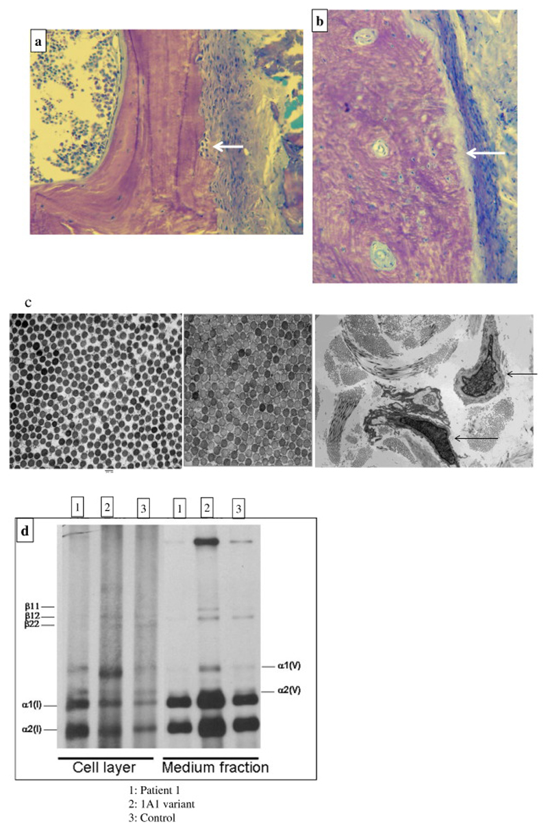
a-b: 3a: Toluidine blue-stained section of an undecalcified trans-iliac bone biopsy, original magnification x400 demonstrating cortex, with periosteum on the right showing high turnover osteopenia with marked sub-periosteal bone resorption (arrow) and normal lamellar bone matrix structure in Patient-1, aged 9 years; 3b: appearance in ‘Classical OI’ with abnormal matrix pattern and increased periosteal bone formation surface (arrow). Toluidine blue; Original magnification of 400x.
c-d: 3c: Electron Microscopy of skin biopsy from Patient 1(left image) compared to normal control (middle image) showing normal collagen in mid-reticular dermis with mildly reduced mean collagen fibril diameter (CFD); Original magnification of 20,000x; Lower magnification (right image) with arrows indicating deep reticular dermal fibroblast with expanded protein filled rough endoplasmic reticulum; Original magnification x2,600.
3d: normal collagen species analysis with no deviation from control (1 and 3) in comparison to patient with a COL1A1 variant (2).

