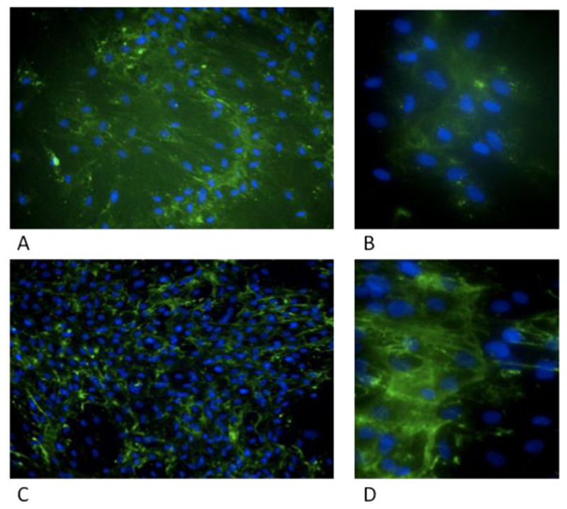
Figure 6.
NBAS patient 1 cultured fibroblasts (A,B) and control sample (C,D) were grown for 3 days in 96 well plates, fixed and stained with anti-Col1A1 antibody (green) and Hoechst (blue) and imaged using a high content microscope. (A, B) show increased diffuse cytoplasmic staining. Collagen bundles from control (C, D).

