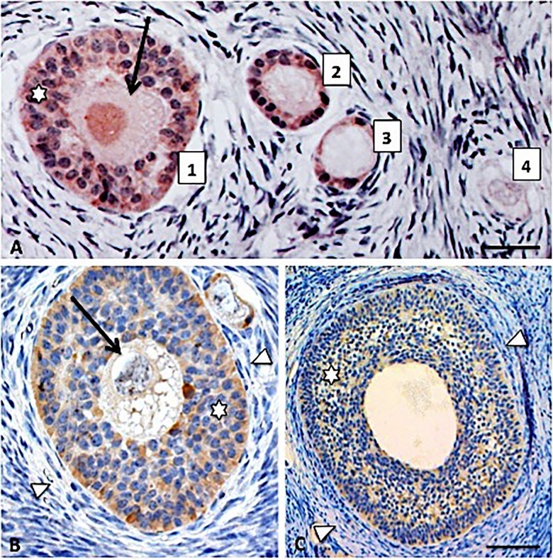
Fig 1.
Representative AMH staining (brown) in the porcine ovary: (A) 1. Small preantral follicle, 2. Primary follicle, 3. Recruited primordial follicle, 4. Quiescent primordial follicle in which AMH staining is absent; (B) Large preantral follicle; (C) Small antral follicle. Oocytes are indicated by arrows, AMH positive granulosa cells by asterisks and theca cells by arrowheads. Scale bars represent 15 μm (A), 30 μm (B), 60 μm (C).

