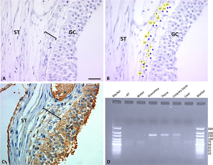
Fig 5. Representative pictures used for LCM capturing of theca cells in preovulatory follicles.
(A) Preovualtory follicle prior to capturing of cells; (B) Same follicle after laser capturing of cells, areas where cells have been captured are indicated by numbered yellow squares; (C) Same follicle stained with the AMH antibody, positive granulosa and theca cells are indicated (brown staining); (D) Agarose gel electrophoresis of AMH cDNA products after RT-PCR (product size—145 bp), lane 1 marker, lane 2—RT (control), lane 3 water (control), lane 4 granulosa cells, lane 5 theca cells, lane 6 luteal cells, lane 7 liver (negative control), lane 8 marker. GC—granulosa layer; black bar—theca layer; ST—stroma. Scale bar represents 30μm (A-C).

