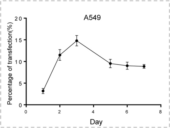
Figure 2b:

Human lung adenocarcinoma cells (A549) were transduced with green fluorescent protein (GFP)/herpes simplex virus thymidine kinase/lentiviral particles. (a) Fluorescent microscopy showed increasing expression of GFP in cells over 7 days. (b) Quantitative assay with flow cytometry enabled confirmation of the level of GFP gene expression (mean ± standard deviation), reaching a peak on day 3 after gene transduction and maintaining a relatively high level over the following days.
