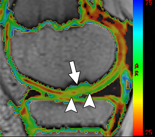
Figure 3c:

MR images in a 13-year-old boy with stable juvenile osteochondritis dissecans (JOCD) lesion of the medial femoral condyle. (a) Sagittal proton density–weighted fast spin-echo and (b) sagittal fat-suppressed T2-weighted fast spin-echo images of the knee show a JOCD lesion on medial femoral condyle (arrows) with surrounding T2-weighted non–fluid-like high-signal rim (arrowhead). (c) Corresponding T2 color map shows diffuse increased T2 color map signal within JOCD lesion (arrow). The cartilage overlying the JOCD lesion has focal area of increased T2 color map signal (arrowheads in c).
