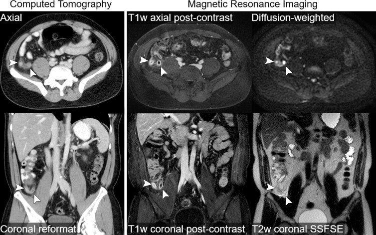
Figure 2:
Intravenous contrast-enhanced CT and intravenous contrast-enhanced MR images in a 41-year-old woman with abdominal pain and uncomplicated appendicitis (arrowheads). Thin axial and reformatted coronal CT images acquired after administration of iodinated contrast material are shown. Selected images from MR imaging protocol are also shown, including coronal and axial postcontrast T1-weighted (T1w) images acquired after administration of gadolinium-based contrast material, a coronal T2-weighted (T2w) single-shot fast spin-echo (SSFSE) image, and an axial diffusion-weighted image. Both CT and MR images accurately depict appendiceal wall thickening, periappendiceal stranding, and mucosal enhancement. The T2-weighted MR image also depicts periappendiceal fluid, and the inflamed appendix is very conspicuous on the diffusion-weighted MR image.

