Figure 2.

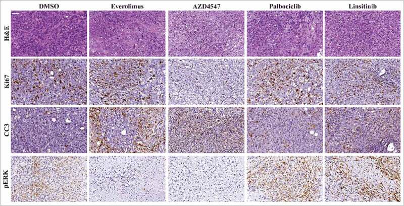
Ex Vivo Organ Culture Histopathology Following Treatment. Representative images from treated tumors. Tumors were stained with H&E, Ki67 proliferation marker, apoptosis marker cleaved-caspase-3 (cc3) and phosphorylated ERK (pERK).

Ex Vivo Organ Culture Histopathology Following Treatment. Representative images from treated tumors. Tumors were stained with H&E, Ki67 proliferation marker, apoptosis marker cleaved-caspase-3 (cc3) and phosphorylated ERK (pERK).