Figure 1.

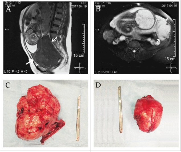
MRI and gross appearance of tumors. (A) MRI showing the dysgerminoma occupying the whole Douglas cul-de-sac. (B) MRI showing the desmoid tumor at the left side of uterus. (C) The right ovarian mass and fallopian tube after excision. (D) The retroperitoneal mass after excision.
