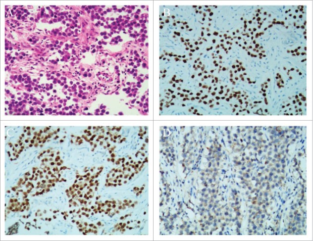
Figure 2.

Histopathological results of the ovarian dysgerminoma and immunohistochemical staining. (A) Histopathology showing sheets of tumor cells, separated by fibrous septa (H & E stain, × 100). (B) SALL-4 staining was positive (SALL-4, × 100). (C) Oct-4 staining was positive (Oct-4, × 100). (D) CK staining was weekly positive (CK, × 100).
