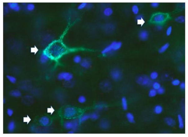
Fig. 2.
Neurons in a section from mouse prefrontal cortex labeled with Wisteria floribunda agglutinin (WFA; green; arrows) to label PNNs and DAPI (blue; to stain cell nuclei). This image demonstrates the extent to which PNNs surround both neuron cell bodies (green staining surrounding DAPI-labeling) and processes.

