Figure 2.
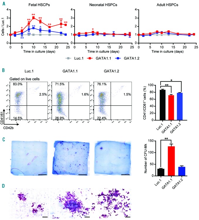
GATA1s mutations in fetal hematopoietic stem and progenitor cells (HSPCs) promote the hyperproliferation of megakaryocytic progenitors. Fetal, neonatal and adult HSPCs were transduced with GATA1-targeting (GATA1.1 and GATA1.2) or control (Luc.1) sgRNAs. (A) Relative number of transduced HSPCs (normalized to the number of Luc.1 cells) grown in liquid cultures supporting megakaryocytic differentiation. Data from 1 of 4 independent experiments performed in replicates are shown as mean±Standard Deviation (SD). *PANOVA<0.05, **PANOVA<0.01. (B) (Left) Representative flow cytometry plots (from 3 independent experiments), showing expression of the common megakaryocytic markers CD41/CD61 and CD42b in fetal HSPCs (pre-gated on live cells). Percentages of each population are indicated. (Right) Percentage of fetal CD41/CD61+ cells on day 10 of megakaryocytic differentiation. Data from 2 independent experiments performed in replicates are shown as mean±SD. *PANOVA<0.05, **PANOVA<0.01. (C) (Left) Photographs (scale bar 5 mm) of CFU-MK assays using lentivirally transduced fetal HSPCs. (Right) Number of CFU-MKs. **PANOVA<0.01. (D) Microscopic images of CFU-MK assays (400× original magnification; scale bar 100 μm).

