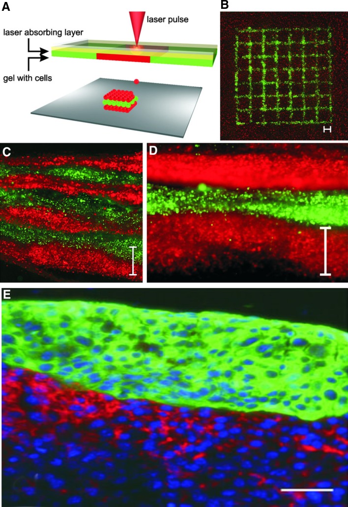
Fig. 3.
Sketch of the laser printing setup a A printed grid structure b of fibroblasts (green) and keratinocytes (red) demonstrates micropatterning capabilities of the laser printing technique. Seven alternating colour layers of red and green keratinocytes c and the magnified view d. Each colour layer consists of four printed sublayers. A histological section was prepared 18 h after printing. Scale bars are 500 µm. In picture e the fibroblasts are stained in red (pan-reticular fibroblast), keratinocytes are stained in green (cytokeratin 14) and cell nuclei are stained in blue (Hoechst 33342). In this case, scale bar is 50 µm. Adopted with permission from (Koch et al. 2012)

