Figure 3.
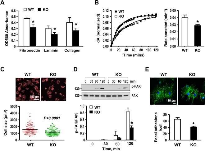
Gal-1 deficiency attenuates VSMC adhesion, spreading and focal adhesion formation. (A) Serum starved WT and Gal-1-KO VSMCs were plated on indicated matrix-coated 96-well plate for 1 h in culture. Nonattached cells were washed out and attached cells were stained with crystal violet and absorbance at 560 nm determined. Data are mean ± SE of 4 independent experiments. *P < 0.05 vs WT cells. (B) Serum-starved WT and Gal-1-KO VSMCs were applied to SPR sensing fibronectin-coated chip cell and the adhesion dynamics recorded for a period of 2 h. The time course plots showing the changes of SPR signal (dA) after normalization with the plateau dA in WT and Gal-1KO VSMCs. The values of rate constant shown are mean ± SE of 4 independent experiments. *P < 0.05 vs WT VSMCs. (C) Serum starved WT and Gal-1-KO VSMCs were plated on fibronectin-coated cover slips for 2 h. Cells were then fixed and stained with rhodamine-conjugated phalloidin. Sizes of spreading WT& Gal-1-KO VSMCs were quantified. (D) Serum-stared WT and Gal-1-KO VSMCs were plated on fibronectin-coated plates for indicated times. Cells were then harvested and FAK phosphorylation was examined by Western blot analysis. The quantitative data were mean ± SE of 3 independent experiments. *P < 0.05 vs WT cells. Uncropped images of immunoblots are shown in Supplementary Fig. 4A. (E) FA formation was examined by confocal immunofluorescence using antibody against vinculin. The FA numbers in WT and Gal-1-KO cells were quantified from 50~70 cells in each group compiled from 3 independent experiments. *P < 0.05 vs WT cells.

