
Figure 12. Responses of cultured preBötC glia to ATP are sensitive to depletion of intracellular calcium stores.
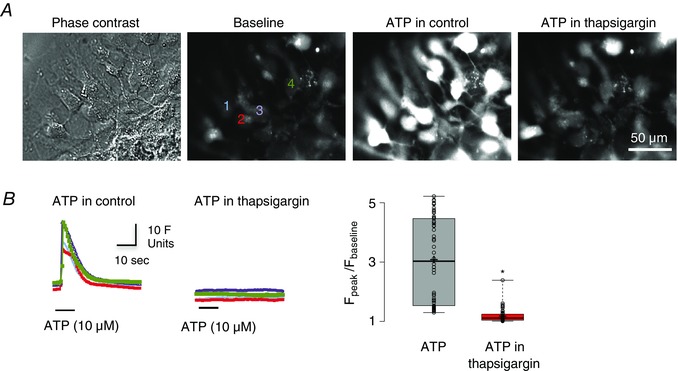
A, images showing a phase contrast image of cultured glia (far left) and fluo‐4 Ca2+ fourescence under baseline conditions (middle‐left), during local application of ATP (10 μm, 10 s) (middle‐right) and during local application of ATP after pre‐application of SERCA inhibitor thapsigargin (50 nm, 30 min) (far right). B, time course of ATP‐evoked fluorescence changes measured from four regions of interest (numbered in A) in control (left) and after thapsigargin application (middle). Group data (n = 54 cells, from four culture plates, each plate was from one animal) showing relative changes in fluorescence (F peak/F baseline) in response to ATP and to ATP in the presence of thapsigargin (right) (n = 54 cells; *significant difference between means P < 0.001, paired t test).