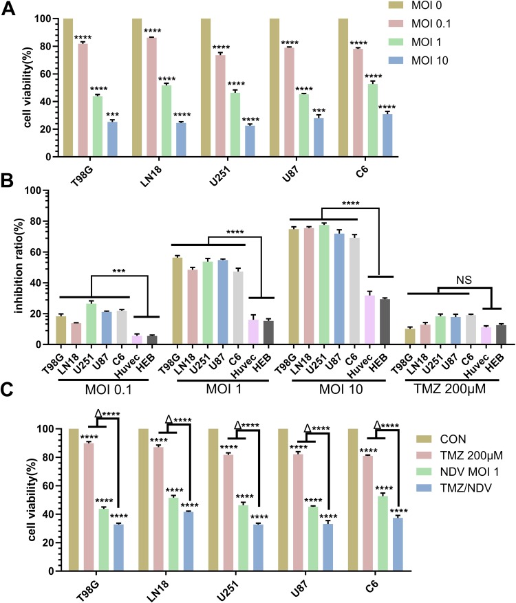
Figure 1.
NDV-LaSota and the combination of TMZ and NDV reduce GBM cell growth in vitro. (A) We infected the T98G, LN18, U251, U87 and C6 cell lines with different MOIs (0, 0.1, 1, or 10) of NDV-LaSota and analyzed cell viability at 36 hours using an MTT assay. The data are presented as the mean ± SD, (*P < 0.01, n = 3, MOI 0.1 vs control, MOI 1 vs MOI 0.1, MOI 10 vs MOI 0.1). (B) Growth inhibition rates after infection of the same strains with the same MOIs as in (A). Every MOI of NDV-LaSota had lower inhibitory effects on HUVECs and HEB cells than on any cancer cell line. TMZ (200 μM) had the same effects on cancer cell lines and normal cell lines (*P < 0.01, n = 3, HUVECs and HEBs vs cancer cell lines). (C) GBM cells were treated with medium, TMZ (200 μM), NDV-LaSota (MOI 1), or a combination of TMZ (200 μM) and NDV-LaSota (MOI 1) (*P < 0.01, n = 3, TMZ and NDV vs control; Δ* P < 0.01, n = 3, TMZ and NDV vs TMZ alone or NDV alone). All results were obtained 36 hours after infection with NDV.

