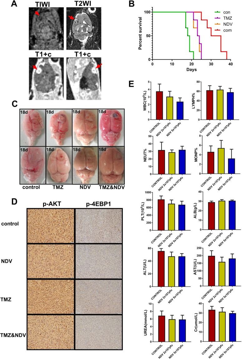
Figure 5.
(A) Eight days after Wistar rats were intracranially injected with C6 cells, creation of the animal models was confirmed by MRI. (B) Survival curves for the animals in the four experimental groups. (C) Eighteen days after intracranial injection of C6 cells, intracranial tumor growth was significantly inhibited in the combination-treated group. (D) Immunohistochemistry analysis of p-AKT(Ser473) and p-4EBP1(Thr37/46) in the implanted tumors. (E) Twenty-four normal rats were intravenously injected with saline, 2 × 10⁷ pfu NDV, or 5 × 10⁷ pfu NDV once every 3 days for a total of three injections, and blood was then collected for routine and biochemical examination. White blood cell and platelet counts differed significantly among the groups, but all values were within the normal ranges. The remaining indices did not differ significantly among the groups (*P < 0.05, n = 6).

