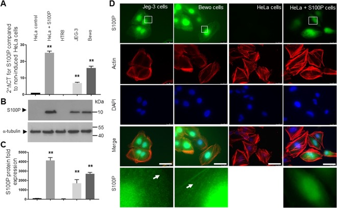
Figure 2.
S100P is expressed in Jeg-3 and Bewo but not HTR8 EV trophoblast cell lines. HTR8, Bewo and Jeg-3 cells, along with HeLa A3 induced for S100P expression (or their non-expressing counterparts), were grown for 48 hours prior to collection for mRNA qPCR analysis (A) or 72 hours prior to collection for protein Western blotting (B). mRNAs were isolated using TRIS reagent followed by reverse transcription and quantitative PCR analysis using S100P and β-actin primers, as described in Methods. Data is presented as 2∆CT mean values ± SD of 3 independent samples of a representative experiment compared to the non-induced HeLaA3 cells. **P < 0.0001 (one way- ANOVA). (A) For the protein levels, cells were collected and solubilised in Laemmeli buffer and equal loading were separated by SDS-PAGE electrophoresis. Western blotting was carried out and membranes probed for S100P or α-tubulin and cropped images are presented. (B) Expression levels of S100P were measured by densitometric analysis, normalised to α-tubulin and presented in comparison to the non-induced HeLa A3 cells as percentage mean values ± SD of 3 independent samples compared to the non-induced HeLa A3 cells. **P < 0.0001 (one way- ANOVA). (C) For immunostaining, Bewo and Jeg-3 cells, along with HeLa A3 induced for S100P expression (or their non-expressing counterparts) were seeded on fibronectin-coated coverslips and grown for 48 hours prior to fixation, permeabilisation and staining for S100P and actin. Cells were mounted and viewed using epifluorescence microscopy. (D) Images in the last row correspond to the focused regions of the highlighted cells. Bar corresponds to 25 μm.

