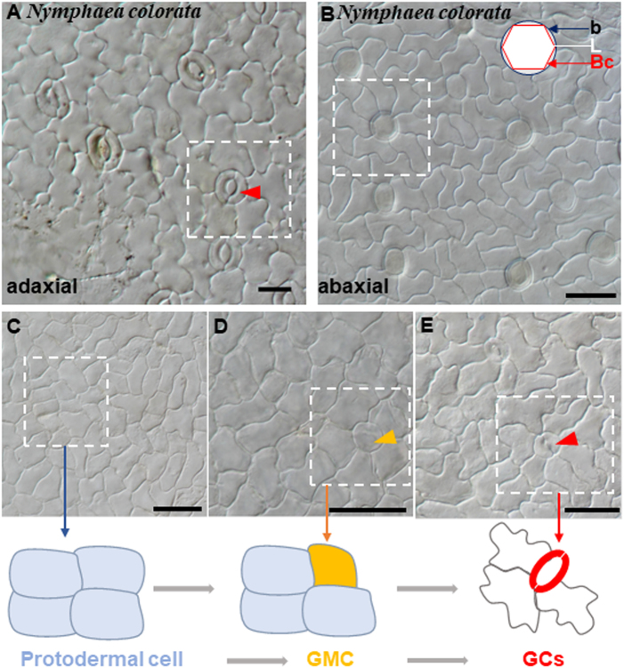
Fig. 1. Stomatal structures and development process in Nymphaea colorata.
a The upper epidermis of N. colorata with anomocytic stomata. b Abaxial hydropote complex structures of N. colorata with base (b) formed by anticlinal contact cell walls, the lens-shaped cell (L), and the bowl-shaped cell (Bc). c-e Micrograph of stomata at different developmental stages in adaxial leaf surfaces. c Squared patterning, a protodermal cell. d Large round cells are putative GMCs (orange arrow). e Stage with maturing stomata (red arrow). Schematic diagram of stomatal development. A protodermal cell (pale blue) that differentiated directly into a guard mother cell (orange); then, the GMC divided into GCs (red)

