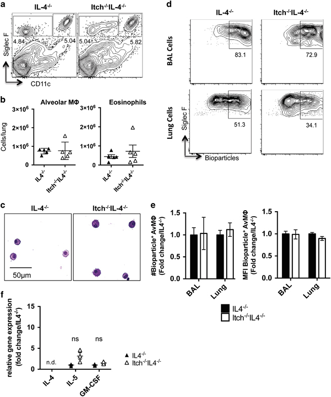
Figure 3.

Numbers of alveolar macrophages are restored in Itch−/− mice lacking IL-4. BAL and lung were collected from uninfected IL-4−/− and Itch−/−IL4−/− mice. (a) Representative flow cytometry plots and (b) quantification of alveolar macrophages and eosinophils in lung cell suspensions. Flow cytometry plots are gated on live, singlet, CD45+. Alveolar macrophages are CD11c+CD11bloSiglecF+, and eosinophils are CD11c−SiglecF+ (n=5, compiled from two independent experiments). (c) Representative BAL cell cytospin slides stained with modified Giemsa stain and visualized at × 40. (d) Representative flow cytometry plots showing uptake of E. coli bioparticles in BAL and lung alveolar macrophages after intranasal administration. (e) Quantification of numbers and mean fluorescence intensity of bioparticle positive BAL and lung alveolar macrophages. n=5–6, data are compiled from two independent experiments. Significance was calculated by two-way analysis of variance. (f) Cytokine gene expression in whole lung homogenate was quantified by QPCR (n=3). Dots represent individual mice. Significance was calculated using an unpaired t-test.
