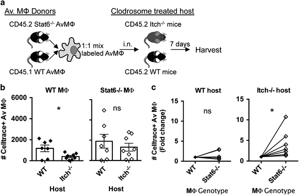
Figure 6.

Stat6−/− alveolar macrophages exhibit improved fitness in the Itch−/− lung environment. (a) Diagram describing experimental design. Alveolar macrophages (AM) were sorted from lungs of donor mice and labeled with cell trace violet, then intranasally transferred to recipient mice that had been pretreated with clodrosomes. Recipient lungs were collected 7 days post transfer. (b) Transferred AM were identified as CD11c+SiglecF+CellTrace violet+. Total numbers of recovered Stat6−/− or WT alveolar macrophages in either WT or Itch−/− host are displayed. (c) The same data from b are displayed in a different way: numbers of Stat6−/− and WT alveolar macrophages detected from the same host are compared in a paired manner. n=7–8, compiled from three independent experiments. Significance was determined by (b) unpaired t-test and (c) paired t-test. * denote P≤0.05, respectively.
