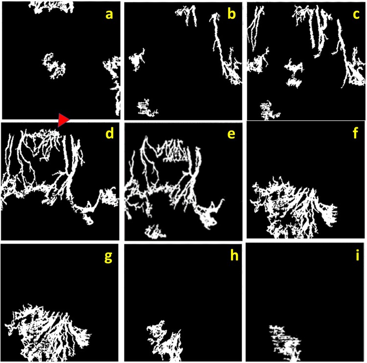
Figure 3.
Sequence of processed ‘en face’ optical coherence tomography angiography (OCTA) demonstrates vessels at different corneal depths. A series of coronal ‘en face’ corneal sections from subepithelial (a) to mid stroma layer (i) of rabbit 5 at week 6, every 20 microns interval, depicting corneal vessels of the same rabbit 5 at week 6 as in Fig. 2. The red Δ indicates the same vessels in area 1 of Fig. 2, which are present in superficial layers (d,e) whereas the fan-like vessels are present in the deeper layers (f,g,h). Note, discontinuity of the vascular network and overlapping of same vessels in different frames caused by unparallel segmentation, which stems from the natural three dimension growth of vessels and steep rabbit corneal curvature.

