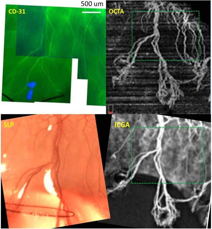
Figure 7.
Example of whole mount cornea showing CD-31 stained blood vessels in suture-induced cornea neovascularization (CoNV) model captured by fluorescence biomicroscopy. A portion of whole mount cornea of rabbit 3 (same as Fig. 1 set 2) shows blood vessels, stained by CD-31. The corresponding area imaged with optical coherence tomography angiography (OCTA), slit-lamp photography (SLP) and indocyanine green angiography (ICGA) images are matched. (Bordered by green dashed squares in OCTA and ICGA). Note, different angle of image acquisition alters position and length of the vessels in each image.

