Fig. 5.
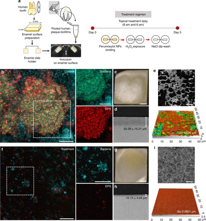
Antibiofilm properties of topical ferumoxytol/H2O2 treatments using an ex vivo biofilm model. a Experimental design and processing. b, Confocal imaging of the morphology of vehicle-control treated biofilm and f biofilm treated with ferumoxytol/H2O2 (white box indicates selected area for close-up images of bacteria and EPS components; Scale bar: 50 µm): total bacteria and S. mutans cells are labelled in blue and green, respectively; EPS are in red (Scale bar: 50 µm). c, Light microscopy images of the enamel surface of untreated biofilm showing “white spot-like caries lesions” and g ferumoxytol/H2O2 treated biofilm showing intact and smooth surface (Scale bar: 1 mm). d, h Lesion depth of the enamel surfaces (control and treated). e, i Representative confocal topography of enamel surfaces and enamel roughness (control and treated) (Scale bar: 10 µm). The data presented as mean ± s.d. from triplicates of two independent experiments (n = 6)

