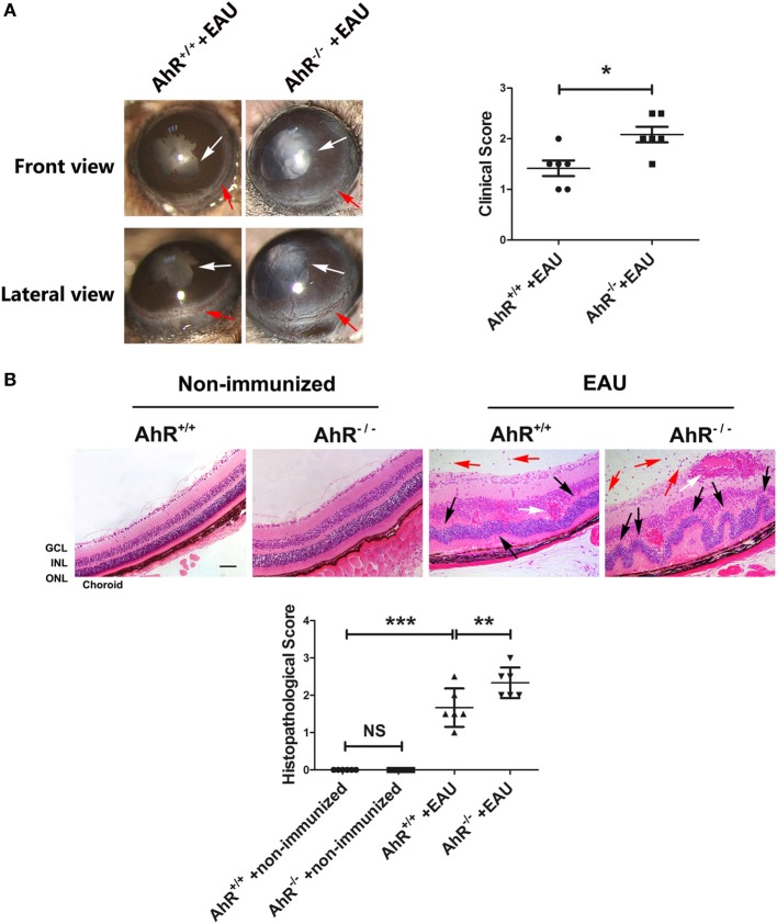
Figure 1.
Effect of aryl hydrocarbon receptor (AhR) knockout on clinical and histological characteristics of experimental autoimmune uveitis (EAU). (A) Left, representative slit-lamp images of AhR−/− mice and AhR+/+ mice after immunization from the frontal and lateral view. White arrow, posterior synechiae. Red arrow, conjunctival hyperemia. Right, quantification of clinical score of AhR−/− mice and AhR+/+ mice after EAU (n = 6/group; mean ± SD; *p < 0.05; unpaired Student’s t-test). (B) Upper, representative hematoxylin and eosin images of eye sections in AhR−/− mice and AhR+/+ mice after EAU induction or without immunization. Abbreviations: GCL, ganglion cell layer; INL, inner nuclear layer; ONL, outer nuclear layer. Black arrow, retinal fold. Red arrow, vitreous infiltration. White arrow, vasculitis. Scale bar, 30 µm. Lower. Quantification of histopathological score of AhR−/− and AhR+/+ EAU mice (n = 6/group; mean ± SD; NSp > 0.05,** p < 0.01; ***p < 0.001; one-way ANOVA).

