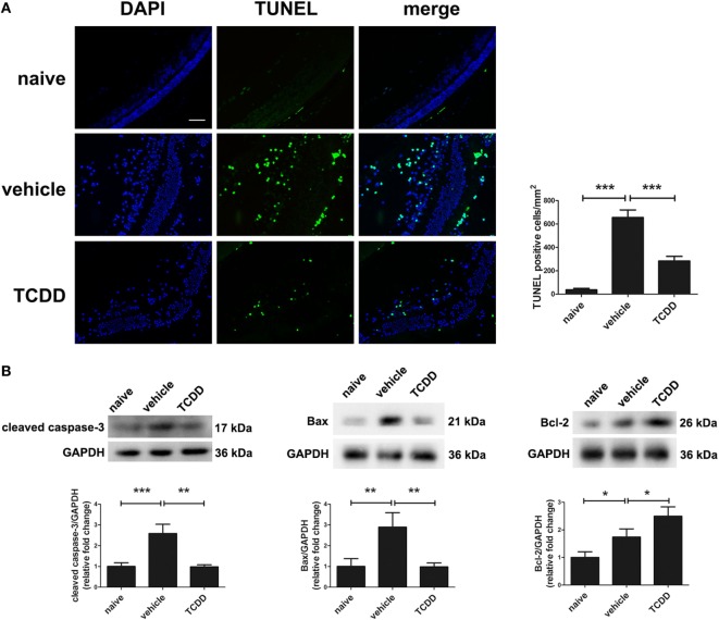
Figure 7.
TCDD treatment decreased apoptotic cell death in the retinas of experimental autoimmune uveitis mice. (A) Left, representative TdT-mediated dUTP nick end labeling (TUNEL) images of eye sections in naive, vehicle, and TCDD-treated mice. Scale bar, 30 µm. Right, quantification of TUNEL-positive cells (n = 4/group; mean ± SD;*** p < 0.001; one-way ANOVA). (B) Upper, representative western blotting images of cleaved caspase-3, Bcl-2 associated X protein (Bax), and Bcl-2 in retinas of naive, vehicle, and TCDD-treated mice. Lower, quantifications of relative fold change of cleaved caspase-3, Bax, and Bcl-2 expressions (n = 4/group; mean ± SD;* p < 0.05; **p < 0.01; ***p < 0.001; one-way ANOVA).

