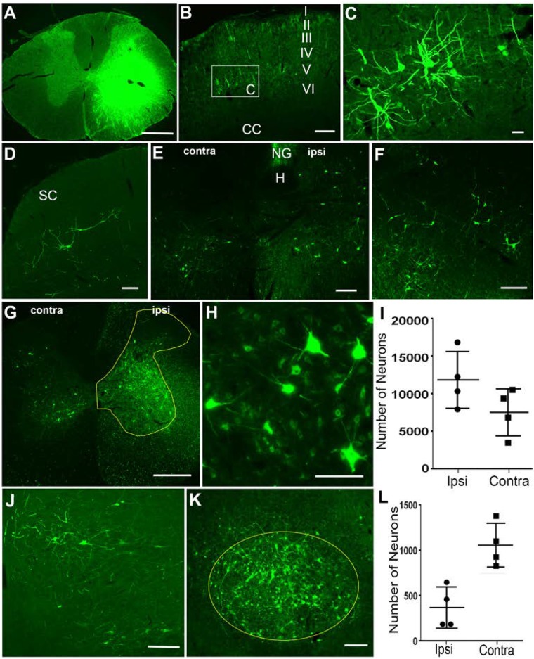
FIGURE 1.
Retrograde labeling of supraspinal and propriospinal neurons after injection of HiRet-GFP into the C6-T1 spinal cord segments. (A) Overexposure of GFP labeling at the HiRet-GFP injection site in the C6 spinal cord showing unilateral expression. (B) Representative image of contralateral somatomotor cortex showing labeling of layer V pyramidal neurons. (C) Higher magnification of box insert of corticospinal neurons from (B). (D) Contralateral superior colliculus (SC) with GFP labeled tectospinal neurons. (E) Caudal medulla showing bilateral GFP-positive neurons within the ventral part of the medullary reticular formation. Hypoglossal nucleus (H); Nucleus Gracilis (NG). (F) GFP-positive neurons labeled within the gigantocellular reticular formation near the ponto-medullary junction. (G) Ipsilateral (ipsi) and contralateral (contra) GFP-labeled propriospinal neurons within the C3-C4 spinal cord segments. GFP positive neurons were quantified from entire ipsilateral (or contralateral) gray matter area within the yellow highlighted region. (H) Higher magnification of GFP-labeled propriospinal neurons ipsilateral to the injection site. (I) Stereological estimates of GFP-positive propriospinal neurons within the C3–C4 spinal cord region showing both ipsilateral and contralateral densities. (J) A few GFP-positive rubrospinal neurons were identified within the ipsilateral red nucleus. (K) However, the vast majority were localized to the contralateral red nucleus. (L) Stereological estimates for the number of GFP-positive rubrospinal neurons identified within either the contralateral or ipsilateral red nucleus. Data is mean ± SD; n = 4. Scale bars: (A,G): 500 μm; (B,D–F,J,K): 200 μm; (C,H): 50 μm.

