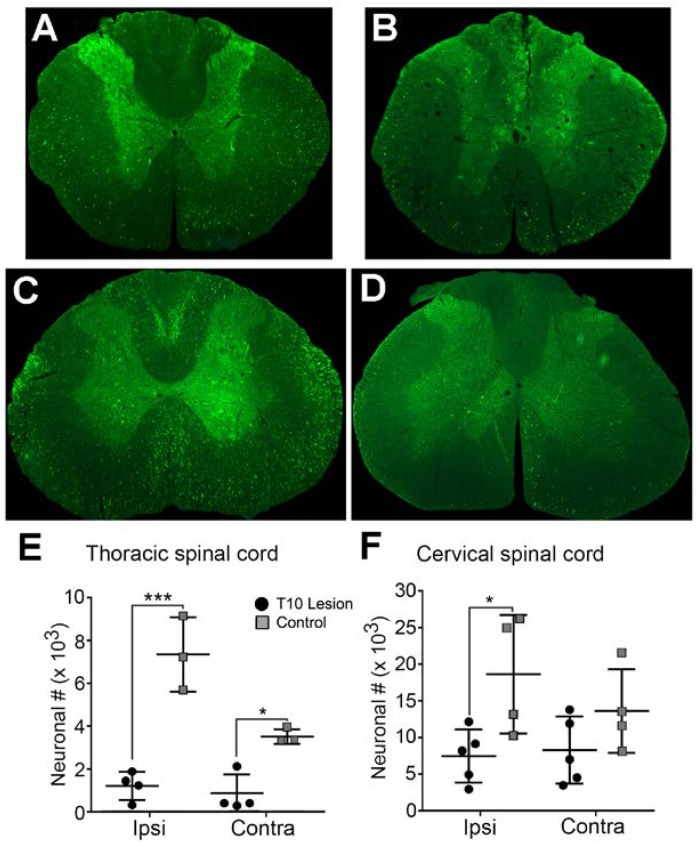
FIGURE 3.
Extent of GFP-expressing neurons within the spinal cord in normal and T10 contused rats. Injection of HiRet-GFP into the non-injured lumbar spinal cord labels many axons and neurons bilaterally within the T7 (A) and C4 (C) regions of the spinal cord. Injection of HiRet-GFP into the lumbar spinal cord 4 weeks after a 200 kD contusion injury to T10 showed a dramatic reduction in the numbers of axons within white matter tracts and neurons within the gray matter at T7 (B) or C4 (D) when compared to the GFP-labeling in normal rats. Stereological estimate of HiRet-GFP expressing neurons between the non-injured (control) and 8 weeks post-injury (lesion) shows a significant reduction in the numbers of bilateral thoracic propriospinal neurons (E) or ipsilateral cervical neurons (F). Comparisons in both graphs by two-way ANOVA [F(1,12) = 9.181, p = 0.0105 (cervical) or F(1,10) = 68.70, p ≤ 0.0001 (thoracic); Sidak multiple comparisons post hoc test, p = 0.0263 (cervical), p < 0.0001 ipsi, p = 0.011 contra (thoracic); ∗p < 0.05, ∗∗∗p < 0.001]. Data are mean ± SD. N = 5 injured, n = 4 control. Scale bars: 500 μm.

