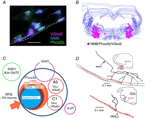
Figure 1. Anatomy of the retrotrapezoid nucleus.

A, cluster of RTN neurons containing mRNA transcripts for VGlut2 and NMB. These neurons are immunoreactive for eGFP, the latter denoting the presence of Phox2b (transverse section, JX‐99 – Phox2b‐eGFP mouse; dashed line identifies the ventral medullary surface (VMS). Scale bar = 25 μm. Unpublished work of R. L. Stornetta. Every neuron is triple‐labelled. B, computer‐assisted mapping of RTN neurons (NMB+) in one mouse. Neurons identified in a one‐in‐three series of 30 μm‐thick transverse sections between 6.65 and 5.63 mm behind bregma. Scale bar = 500 μm (from Shi et al. 2017, with modifications). C, Venn diagram representing the various cell populations located in the parafacial region of the mouse. RTN neurons are defined as positive for Phox2b, VGlut2 and NMB. A least 90% of these neurons contain putative proton sensor TASK‐2 and ∼82% contain GPR4 (Shi et al. 2017). D, location and dendritic structure of two RTN neurons from rat recorded and labelled juxtacellularly in vivo. Note presence of extensive dendrites in the marginal layer (ML) regardless of the position of the cell body (adapted from Mulkey et al. 2004).
