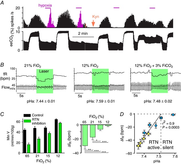
Figure 3. RTN‐dependent and ‐independent activation of breathing by the carotid bodies.

A, single‐unit recording evidence that carotid body stimulation can activate RTN. This RTN neuron was activated by hypercapnia (eeCO2: end‐expiratory CO2) and by brief hypoxia (magenta). Administration of kynurenate i.c.v. (orange arrow), a blocker of excitatory glutamatergic synapses, eliminated the effect of hypoxia selectively whereas the effect of hypercapnia, which is primarily mediated by cell‐autonomous and paracrine effects of CO2, persisted (redrawn from Mulkey et al. 2004). B, evidence that the carotid bodies activate breathing via a pathway that bypasses RTN. Bilateral opto‐inhibition of RTN neurons (conscious rats) reduces breathing under normoxia (21% ) but has no effect under hypoxia (12% ). Breathing rate and amplitude increased under hypoxia despite RTN being silent, demonstrating that the CBs activated breathing via a pathway that bypasses RTN. The addition of a small amount of CO2 restored the breathing reduction caused by RTN inhibition, suggesting that hypoxia‐induced hyperventilation had inhibited RTN via the resultant respiratory alkalosis. Average plasma pH identified in 6 rats is shown below the representative examples (modified from Basting et al. 2015). C, breathing reduction elicited by bilateral optoinhibition of RTN is reduced under hypoxia. Left panel, minute volume before (black bars) and during RTN inhibition (green bars); right panel change in breathing frequency elicited by RTN inhibition. At 12% , breathing is activated but RTN no longer contributes to breathing (right panel). At 15% breathing is unchanged from room air but, as indicated in the right panel, a much smaller portion of the respiratory drive originates from RTN (modified from Basting et al. 2015). D, relationship between arterial pH and effect of RTN inhibition on breathing frequency (f R) and plasma pH was manipulated with hypoxia. Respiratory alkalosis was compensated by adding CO2 (blue symbols) or by inducing metabolic acidosis (acetazolamide, yellow symbols). Above pHa 7.5, RTN inhibition has no effect on breathing consistent with single‐unit evidence that the neurons are silent (e.g. panel B). Below pH 7.5, the breathing reduction elicited by RTN inhibition (change in breathing frequency is depicted) is a linear function of arterial pH, consistent with single‐unit evidence (not shown) that RTN neurons are increasingly active (modified from Basting et al. 2015).
