
FIGURE 2.
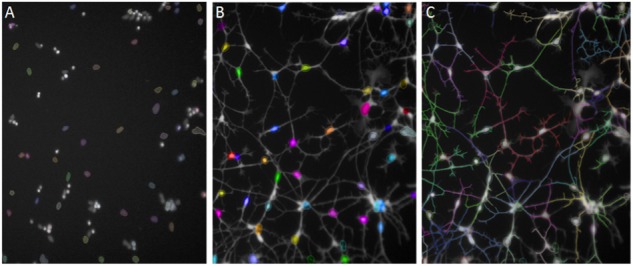
Automated Neuronal Profiling by imaging software at 20× magnification. (A) Nuclei are selected based on size and intensity in Hoechst channel. (B) Nuclei selected in Step A are located in the Calcein AM channel as cell bodies and selected in color by the software. (C) Neuronal processes extending from the cell bodies with Calcein AM stain are traced and the total length of the neurites per cell are measured in numbers of pixels.