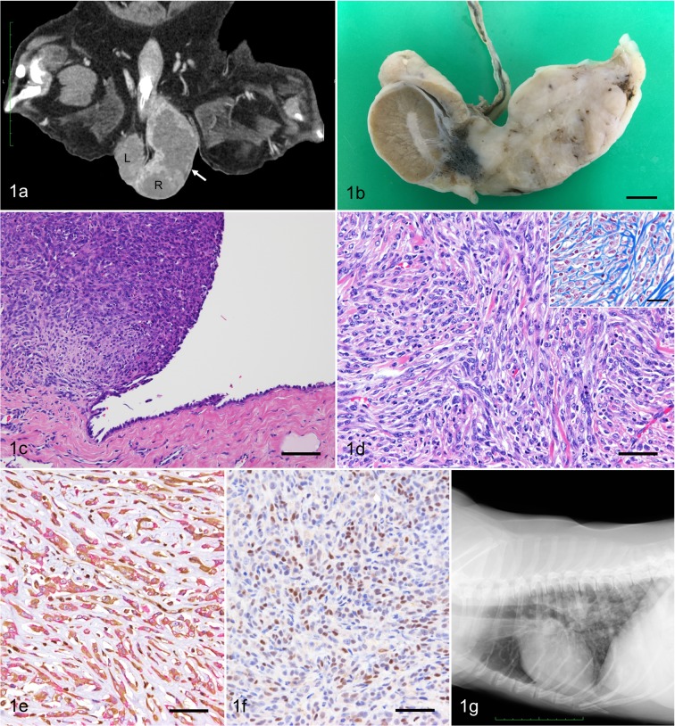
Fig. 1.
(a) Dorsoventral CT view of the scrotum. A mass (7.0 × 4.0 × 3.0 cm, arrow) locates cranial to the right testis (R) in the scrotum. The left testis (L) is intact. (b) Gross appearance of the right testis cut surface shows a white solid mass infiltrating the head of epididymis. Bar=1 cm. (c) The mass is continuous to the mesothelium of the tunica vaginalis testis. HE. Bar=100 µm. (d) Neoplastic cells are spindle-shaped with round to elongate nuclei and scant cytoplasm. HE. Bar=50 µm. Bundles of collagen separates neoplastic cells (inset). Masson’s trichrome stain. Bar=25 µm. (e) The cytoplasm of the tumor cells are positive for both vimentin (brown) and cytokeratin (red). Double immunohistochemistry. Bar=50 µm. (f) Nuclei of the tumor cells are positive for WT-1. Immunohistochemistry. Bar=50 µm. (g) Thoracic radiograph shows multiple nodular lesions with variable size in the lung.

