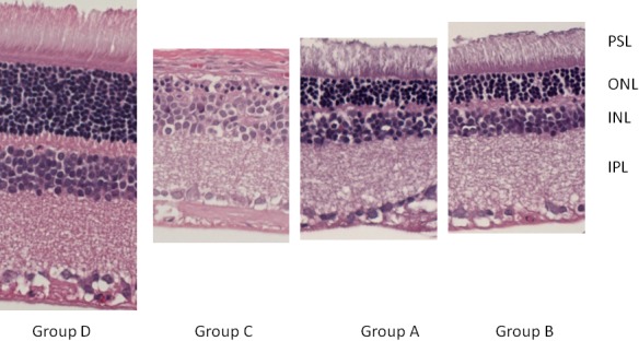
Fig. 1.

Histopathology thickness of retina. The section was excised from the central retina. The ONL and PSL were almost disappeared in Group A, B, C. The loss of OPL and IPL especially in Group C makes ONL and INL barely seen. Group A and B seems to preserve more layer of cells than Group C. IPL=inner plexiform laye, OPL=outer plexiform layer, PSL=photoreceptor segment layer, ONL=outer nucleus layer, INL=inner nucleus layer.
