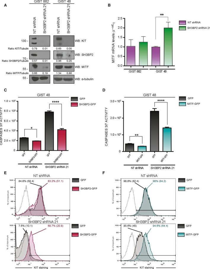
Figure 3.

Microphthalmia‐associated transcription factor expression is reduced in SH3BP2‐silenced cells. SH3BP2 or MITF reconstitution restores cell survival. (A) GIST882 and GIST48 cells were transduced with either control NT (Nontarget) shRNA or SH3BP2 shRNA. Cell lysates were analyzed for MITF protein levels. Tubulin was used as loading control. (B) Real‐time PCR was performed using specific probes against MITF. Statistical significance (**P < 0.01; unpaired t‐test; n = 3; mean ± SEM) is relative to NT shRNA. (C–F) GIST48 cells transduced with either control NT (nontarget) shRNA or SH3BP2 shRNA were posteriorly reconstituted with GFP, SH3BP2‐GFP, or MITF‐GFP. Evaluation of caspase 3/7 activity was performed for SH3BP2 reconstitution (C) or MITF reconstitution (D). Statistical significance (*P < 0.05, ****P < 0.0001; unpaired t‐test; n = 3; mean ±SEM) is relative to GFP‐transduced cells in each case. KIT surface expression by flow cytometry was assayed in SH3BP2‐reconstituted (E) and MITF‐reconstituted (F) SH3BP2‐silenced cells. Percentage of KIT expression and mean fluorescence, in parentheses, are indicated in the FACS histograms.
