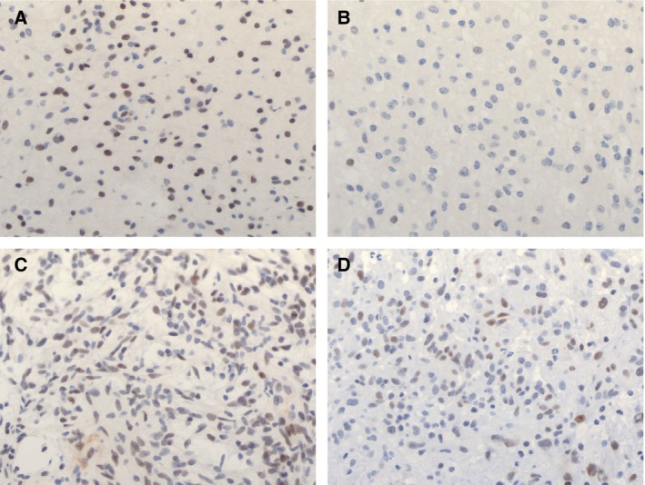
Figure 5.

Immunohistochemical staining for NR2E1 in tumours from three different locations. (A) Dense region of staining seen within an infratentorial tumour and (B) sparse staining within the same tumour. (C) Staining pattern in a midline tumour. (D) Staining pattern in a cortical tumour. Results showed variable expression across tumours with no clear differences between tumours from different locations. Furthermore, there was considerable variation in expression within individual tumour tissues.
